*31-year-old male amateur footballer presented with the complain of pelvic pain for last six months.


What is the most likely diagnosis?
Answer
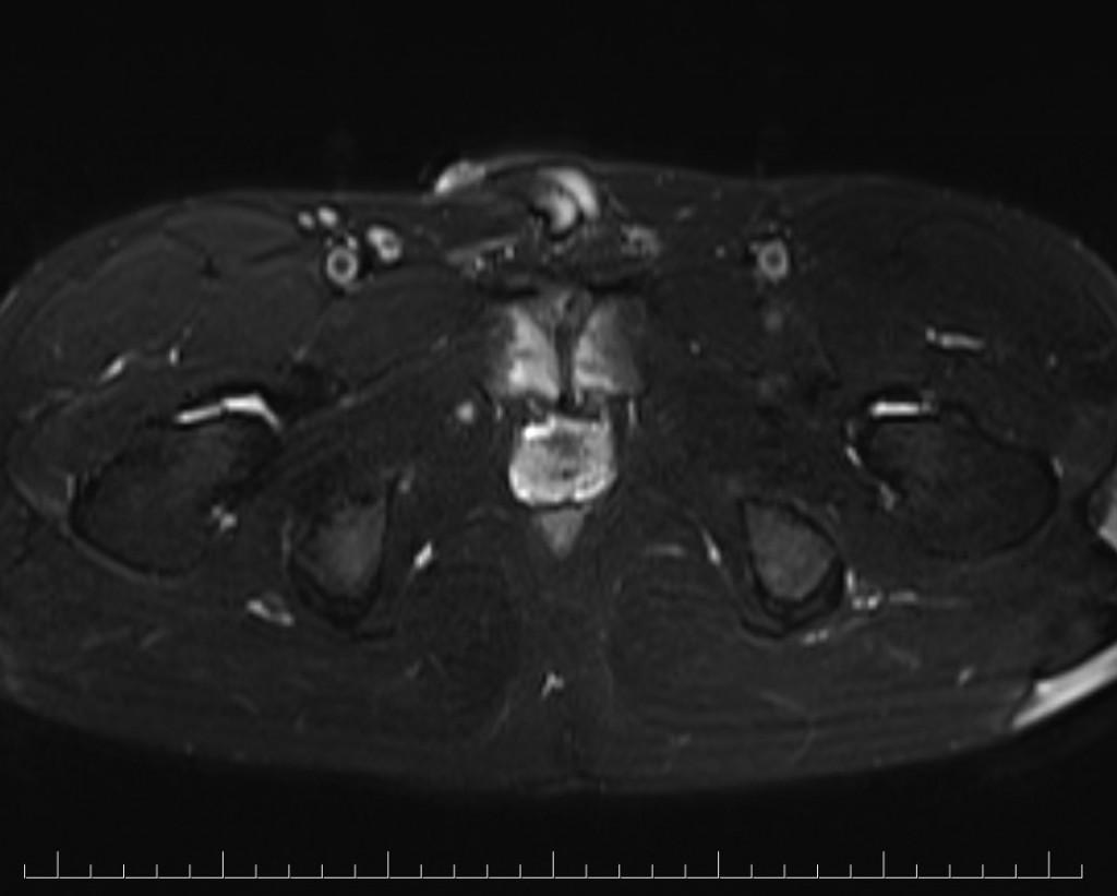
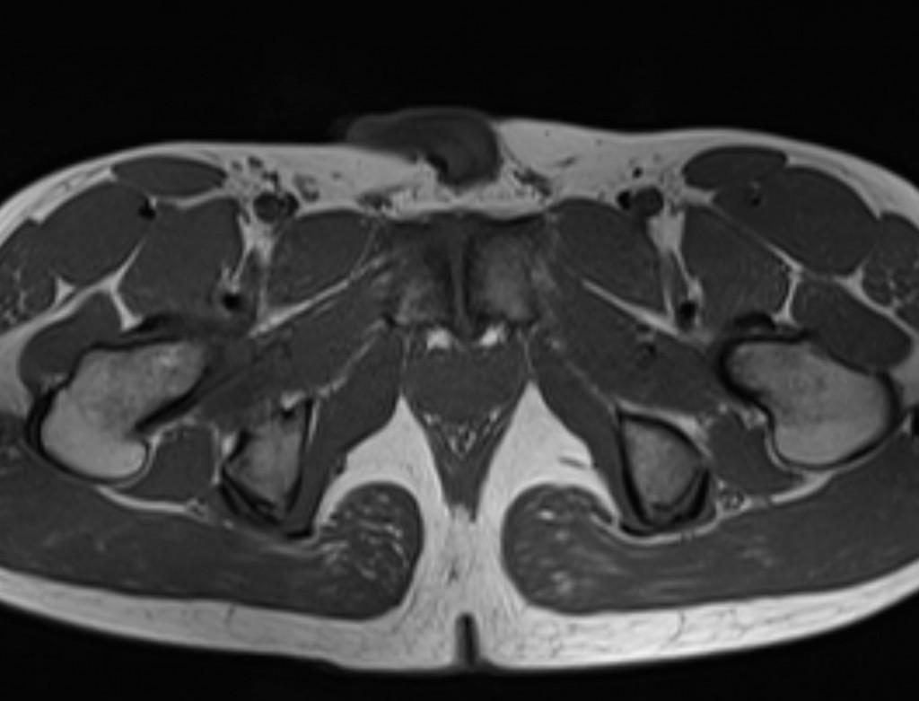
Answer: Osteitis pubis
Case Discussion:
MR images show bone marrow edema and bony margin irregularities (arrows).

Osteitis pubis is a noninfectious inflammation of the symphysis pubis. Clinical presentation is typically with varying degrees of pelvic pain.
Causes
• Pregnancy/childbirth
• Gynecologic surgery
• Urologic surgery
• Athletic activities
• Major trauma
• Repeated minor trauma
• Rheumatological disorders
• Unknown etiologies (most common)
X-ray findings will appear after 4 weeks of symptom onset. Osteitis pubis can be identified with an X-ray. Findings are widening of the pubic symphysis, subchondral erosive change, joint irregularity and sclerosis, and eventually ankylosis. Similar change is also demonstrated with Computed Tomography (CT), but the multi-planar nature of CT has a higher sensitivity than X-ray film.
MRI can reveal bone marrow edema even in asymptomatic individuals. Symphyseal fluid and peripubic soft-tissue edema can also be seen during initial stages. Subchondral sclerosis, subchondral resorption and bony margin irregularities, and osteophytes can be seen with the chronicity of disease.
Differential diagnosis
• Infection
• Hyperparathyroidism
• Osteomyelitis
References:
1. Gibbon WW, Hession PR. Diseases of the pubis and pubic symphysis: MR imaging appearances. AJR Am J Roentgenol. 1997;169 (3): 849-53.
2. Lentz SS. Osteitis pubis: a review. Obstet Gynecol Surv. 1995;50 (4): 310-5.
3. Zoga AC, Kavanagh EC, Omar IM et-al. Athletic pubalgia and the “sports hernia”: MR imaging findings. Radiology. 2008;247 (3): 797-807.
4. Kunduracioglu B, Yilmaz C, Yorubulut M et-al. Magnetic resonance findings of osteitis pubis. J Magn Reson Imaging. 2007;25 (3): 535-9.