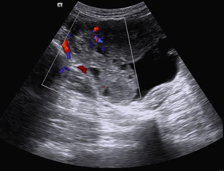
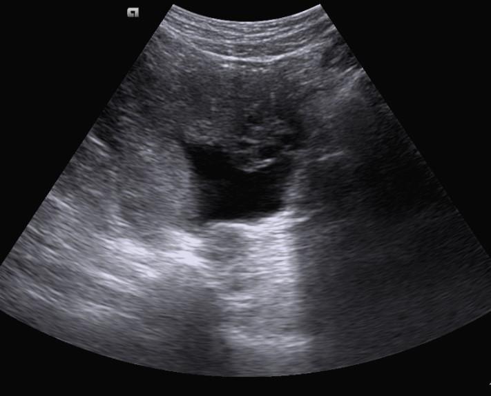
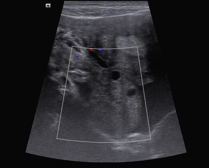
*23-year-old female with right lower quadrant pain.


What is the most likely diagnosis?
Answer
Answer: Ovarian torsion
Case Discussion:
Ultrasound and Doppler images revealed an enlarged hyperechoic right ovary, peripherally displaced follicles, no intra-ovarian venous and arterial flow, and free pelvic fluid.
Ovarian torsion (also sometimes termed adnexal torsion) is a rare but significant cause of acute lower abdominal pain in women. It is refers to rotation of the ovary and portion of the fallopian tube on the supplying vascular pedicle (also termed tubo-ovarian torsion). This can be result in venous, arterial and lymphatic stasis. It is a gynecological emergency and requires urgent surgical intervention to prevent ovarian necrosis.
Ultrasound and Doppler findings
• enlarged hypo-hyperechoic ovary
• peripherally displaced follicles with hyperechoic central stroma
• midline ovary
• free pelvic fluid (>80%)
• an underlying ovarian lesion can be seen
• a long-standing infarcted ovary may have a more complex appearance with cystic or haemorrhagic degeneration
• little or no intra-ovarian venous flow (common)
• absent arterial flow (less common)
• absent or reversed diastolic flow
• normal vascularity does not exclude intermittent torsion (This can occasionally be found due to dual supply from both the ovarian and uterine arteries).
• whirlpool sign of twisted vascular pedicle
Differential diagnosis
• pelvic inflammatory disease
• massive ovarian edema
• oophoritis
References:
1. Bider D, Mashiach S, Dulitzky M et-al. Clinical, surgical and pathologic findings of adnexal torsion in pregnant and nonpregnant women. Surg Gynecol Obstet. 1991;173 (5): 363-6.
2. Dähnert W. Radiology Review Manual. Hubsta Ltd. (2007) ISBN:0781766206.
3. Lee EJ, Kwon HC, Joo HJ et-al. Diagnosis of ovarian torsion with color Doppler sonography: depiction of twisted vascular pedicle. J Ultrasound Med. 1998;17 (2): 83-9.
