*30-year-old pregnant woman with headache and diplopia.
What is the most likely diagnosis?
Answer
Answer: Pituitary apoplexy
Case Discussion:
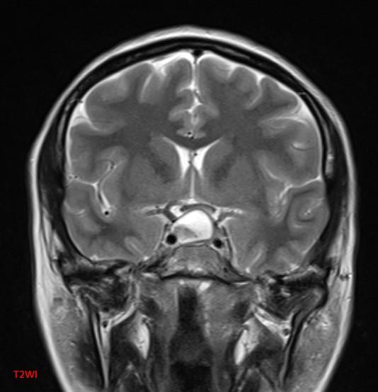
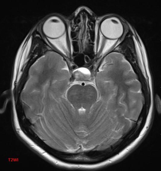
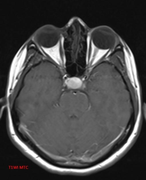
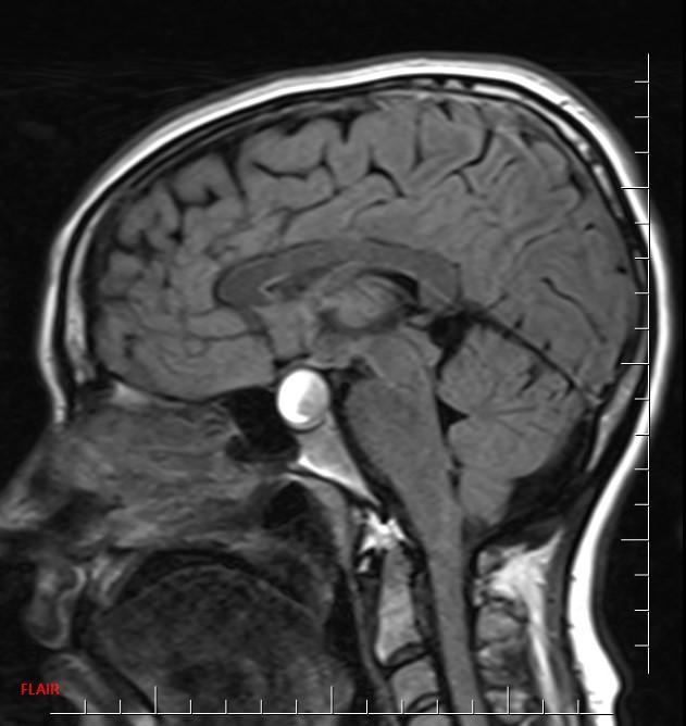
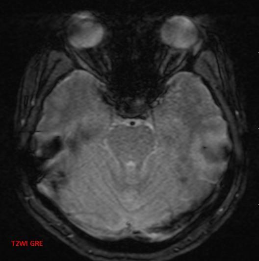
MR images reveal an ovoid lesion (red arrows) in the pituitary fossa with suprasellar extension. The optic chiasm is compressed (yellow arrows). The lesion is T1 and T2 hyperintense showing blood-blood fluid level (blue arrows).

Pituitary apoplexy is a condition due to infarction or haemorrhage of pituitary gland. It is characterized by a sudden onset of headache, visual symptoms, altered mental status, and hormonal dysfunction (1).
MRI typically demonstrates a pituitary region mass. The imaging characteristics of blood on MRI are variable and change with the age of the blood (2, 3).
• T1: variable signal.
• T2: variable signal.
• T1 C+: enhancement variable; usually peripheral.
• DWI: restricted diffusion can be present in solid infarcted components
Differential diagnosis
• Necrotic/haemorrhagic pituitary macroadenoma
• Craniopharyngioma
• Rathke cleft cyst
• Dermoid/teratoma
References:
1. Nawar RN, AbdelMannan D, Selman WR, Arafah BM. Pituitary tumor apoplexy: a review. J Intensive Care Med. 2008;23(2):75-90.
2. Bradley WG. MR appearance of hemorrhage in the brain. Radiology. 1993;189 (1):15-26.
3. Rogg JM, Tung GA, Anderson G et-al. Pituitary apoplexy: early detection with diffusion-weighted MR imaging. AJNR Am J Neuroradiol. 2002;23(7):1240-5.