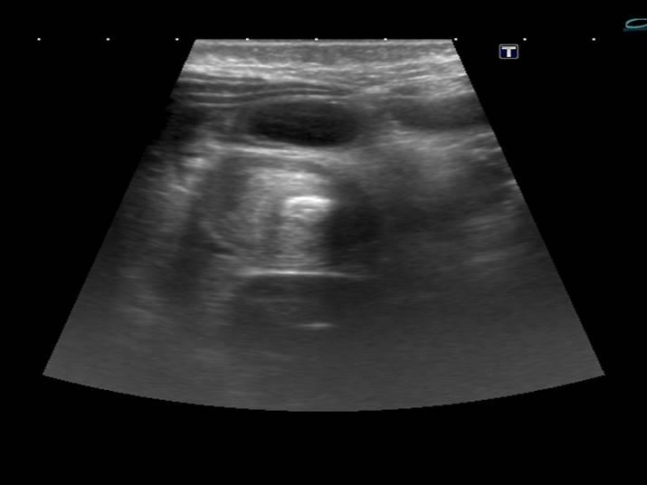
*8 month old female with vomiting and blood in her stool.


What is the most likely diagnosis?
1. Appendicitis
2. Intestinal lymphoma
3. Intestinal lipoma
4. Intussusception
Answer
Answer: Intussusception
Case Discussion:
The AP supine abdominal plain film shows distended loops of gas filled small bowel. Ultrasound images show a target sign-characteristic for intussusception and distended loops of fluid filled small bowel and free fluid between intestinal loops.
Intussusception is a process in which a part of the intestine invaginates into another section of intestine, causing bowel obstruction (1). It is common in children (95%), particularly after the first three months. With early diagnosis and therapy, the mortality rate in children is less than 1%. If untreated, this condition is fatal at high rates in 2-5 days (2).
Intussusception can be caused by a gastrointestinal malignancy (most common cause in adults), benign neoplasms, congenital (Meckel diverticulum, duplication cyst, ectopic pancreas), inflammatory (periappendicitis), trauma (mural haematoma) (2-4).
It can occur anywhere from the duodenum to the rectum, although in children there is a strong predilection for the ileocolic region.
Abdominal plain film may demonstrate an soft tissue mass with a bowel obstruction proximal to it. Ultrasonography is the gold standard in the diagnosis of intussusception. Hallmarks of ultrasonography include the target and pseudokidney signs. Contrast enema is the traditional and most reliable way to make the diagnosis of intussusception in children.
References:
1. Gylys, Barbara A. and Mary Ellen Wedding (2009), Medicasl Terminology Systems, F.A. Davis Company
2. Choi SH, Han JK, Kim SH et-al. Intussusception in adults: from stomach to rectum. AJR Am J Roentgenol. 2004;183 (3): 691-8.
3. Sparnon AL, Little KE, Morris LL. Intussusception in childhood: a review of 139 cases. Aust N Z J Surg. 1984;54 (4): 353-6.
4. del-Pozo G, Albillos JC, Tejedor D. Intussusception: US findings with pathologic correlation–the crescent-in-doughnut sign. Radiology. 1996;199 (3): 688-92.
