*58-year-old male with loss of consciousness.
What is the most likely diagnosis?
Answer
Answer:
Herpes simplex (HSV) encephalitis
Case Discussion:
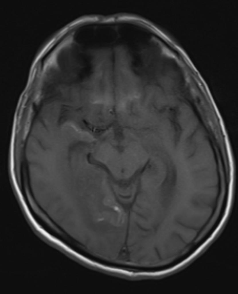
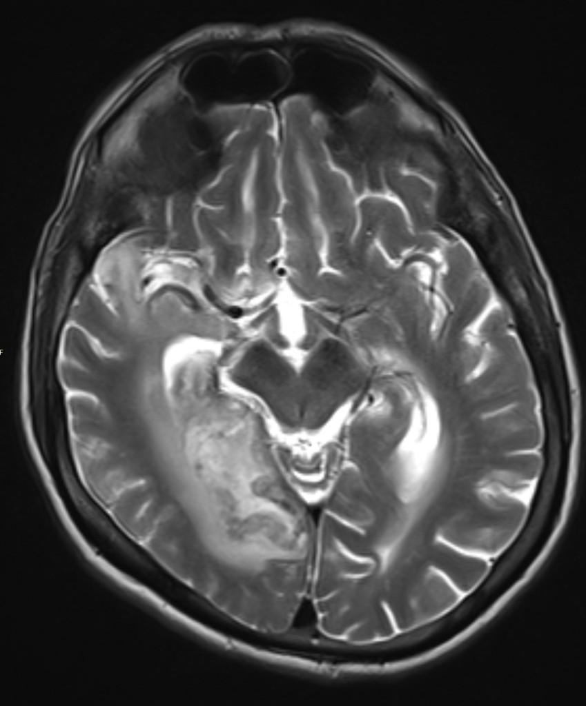
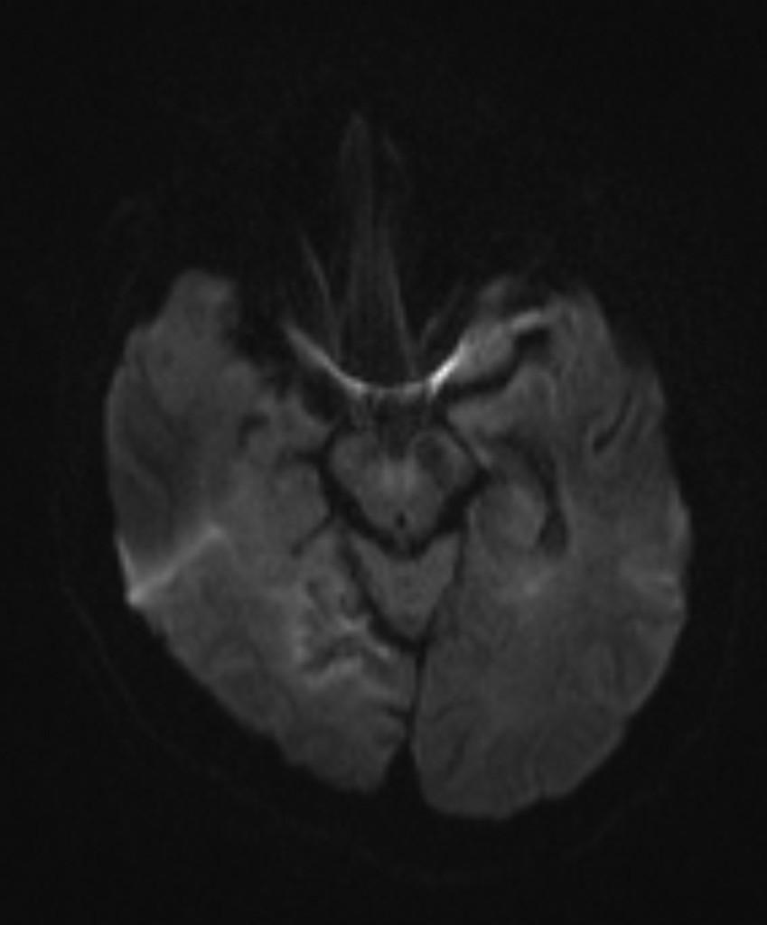
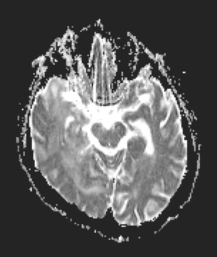
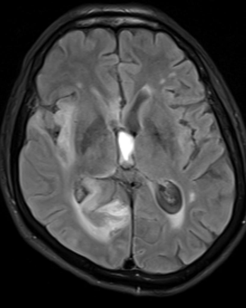
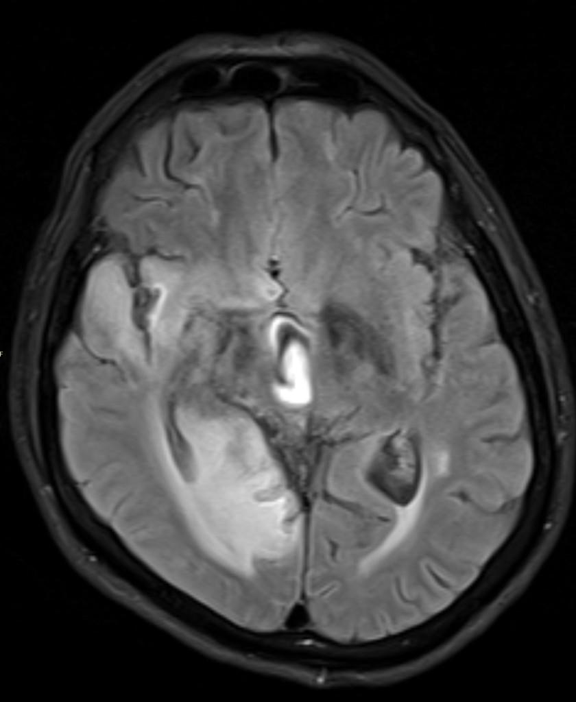
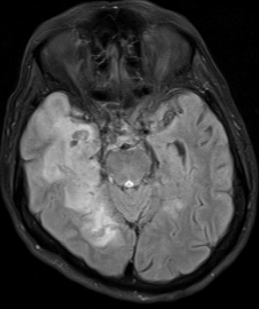
MR images of brain revealed the T2 and FLAIR high signal at the right temporal lobe, along with gyriform and leptomeningeal patterns of enhancement and foci of hemorrhage.
HSV encephalitis is the most common cause of fatal sporadic fulminant necrotising viral encephalitis and has characteristic imaging findings.
Two subtypes are recognised which differ in demographics, virus, and pattern of involvement. They are neonatal herpes encephalitis and childhood-adult herpes encephalitis.
References:
1. Leonard JR, Moran CJ, Cross DT et-al. MR imaging of herpes simplex type 1 encephalitis in infants and young children: a separate pattern of findings. AJR Am J Roentgenol. 2000;174 (6): 1651-5.