*50-year-old female present with left lower quadrant pain for 3 days.
What is the most likely diagnosis?
Answer
Answer: Epiploic Appendagitis
Case Discussion:
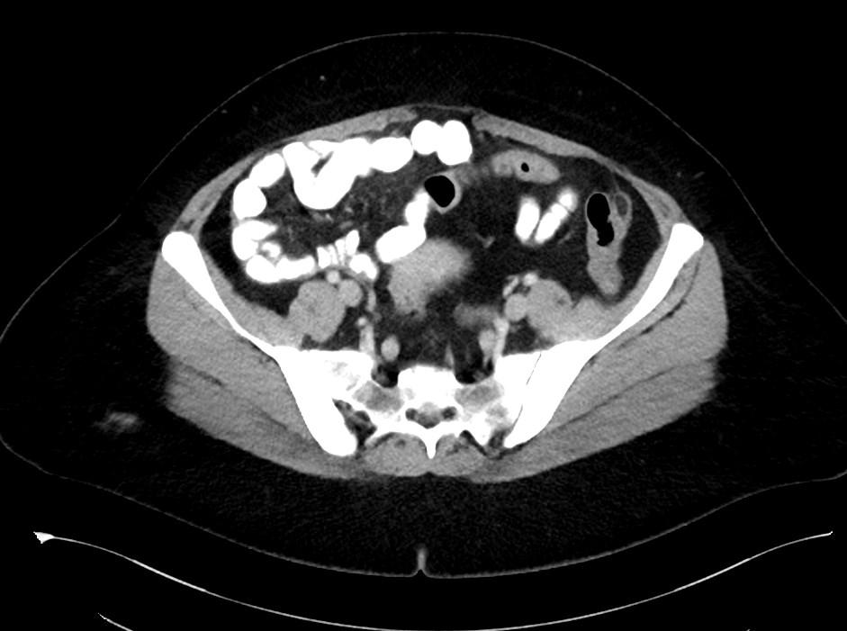
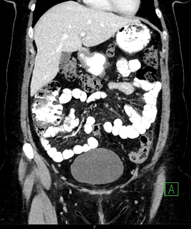
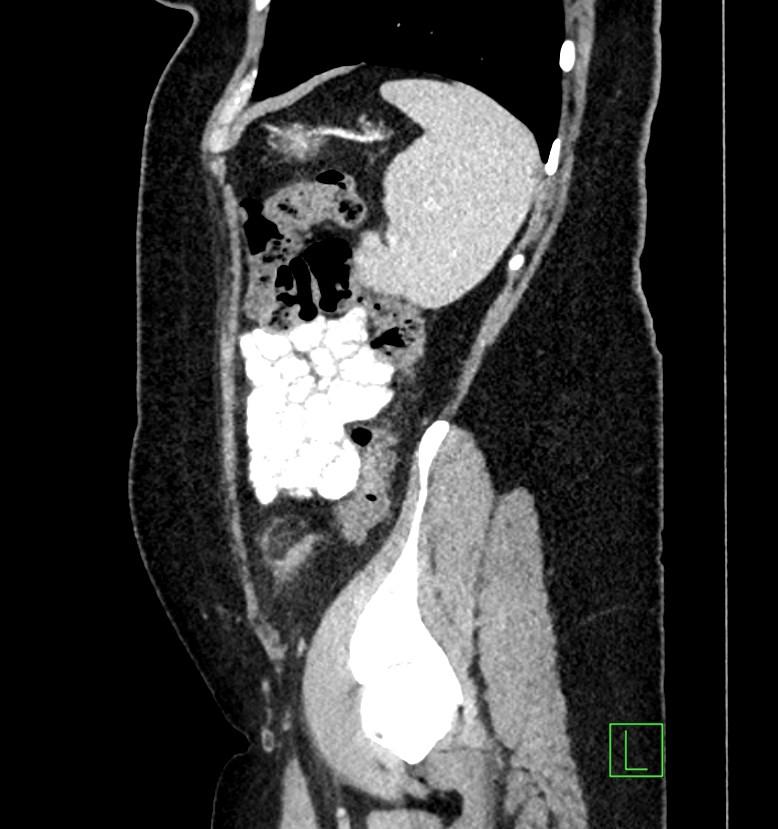
MDCT images showed fatty density lesion adjacent to descending colon contains a characteristic hyperdense ring corresponding to thickened visceral peritoneal lining.
Epiploic appendages are small fatty peritoneal projections from the serosal surface of the colon. The inflammation of epiploic appendages can be due to torsion or venous occlusion. The disease is a self-limiting disease. Sonography and CT demonstrate an inflamed fatty lesion adjacent to the colon, including a characteristic hyperdense ring of thickened visceral peritoneal lining on CT [1].
Omental infarction has a clinical presentation similar to that of epiploic appendagitis. However, imaging illustrates an inflamed fatty mass, larger than in epiploic appendagitis and lacking a hyperdense ring on CT [1].
References:
1. Breda Vriesman AC, Puylaert JBCM.Epiploic appendagitis and omental infarction: pitfalls and look-alikes.Abdom Imaging 2002; 27:20-28