*45-year-old male present with acute confusion, ataxia, and ophthalmoplegia.

What is the most likely diagnosis?
Answer
Answer:
Wernicke encephalopathy
Case Discussion:
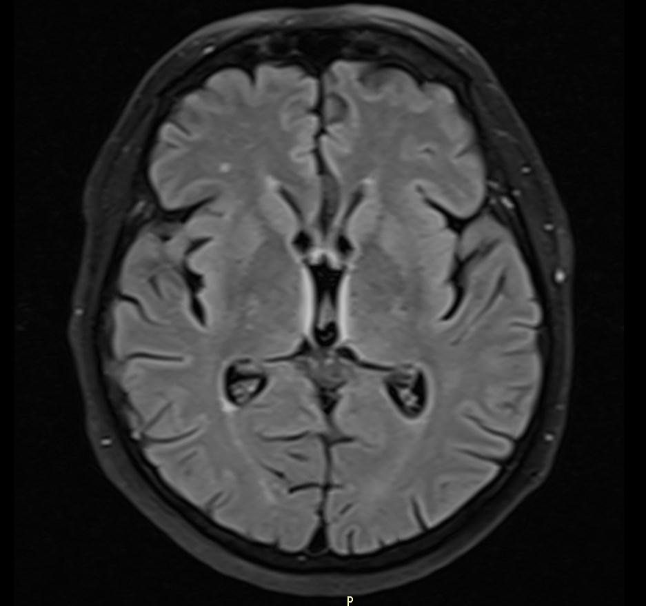
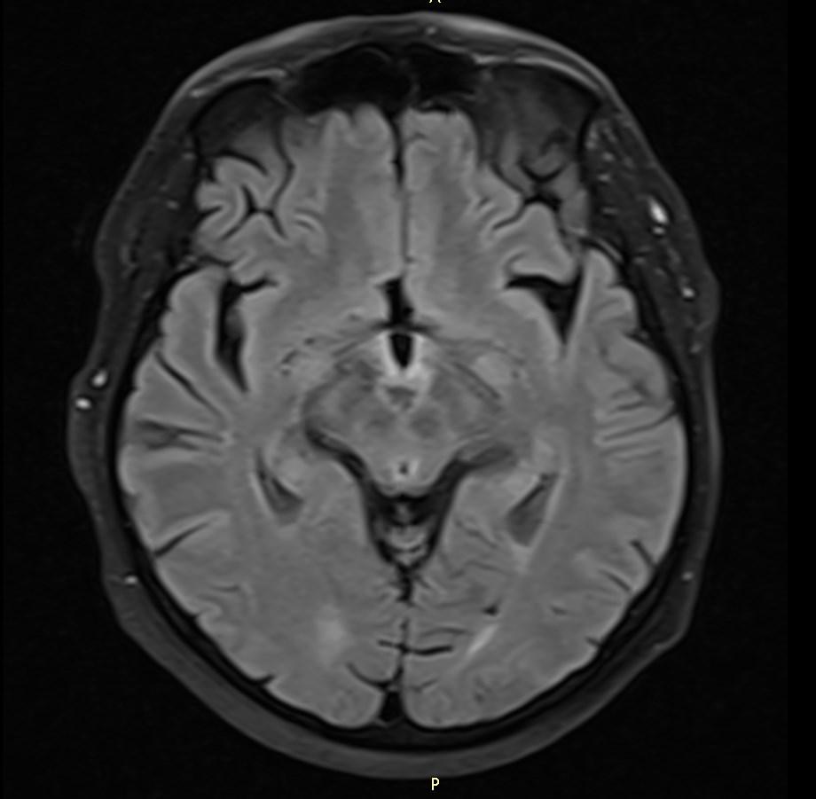
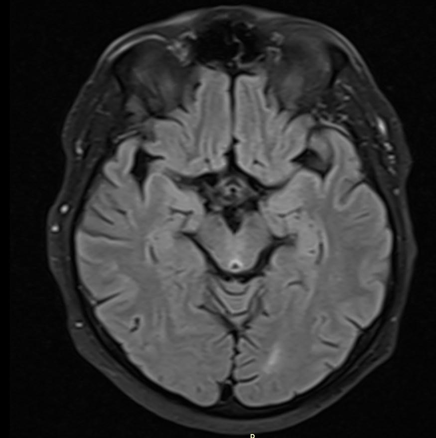
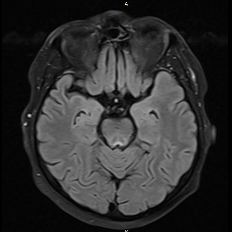
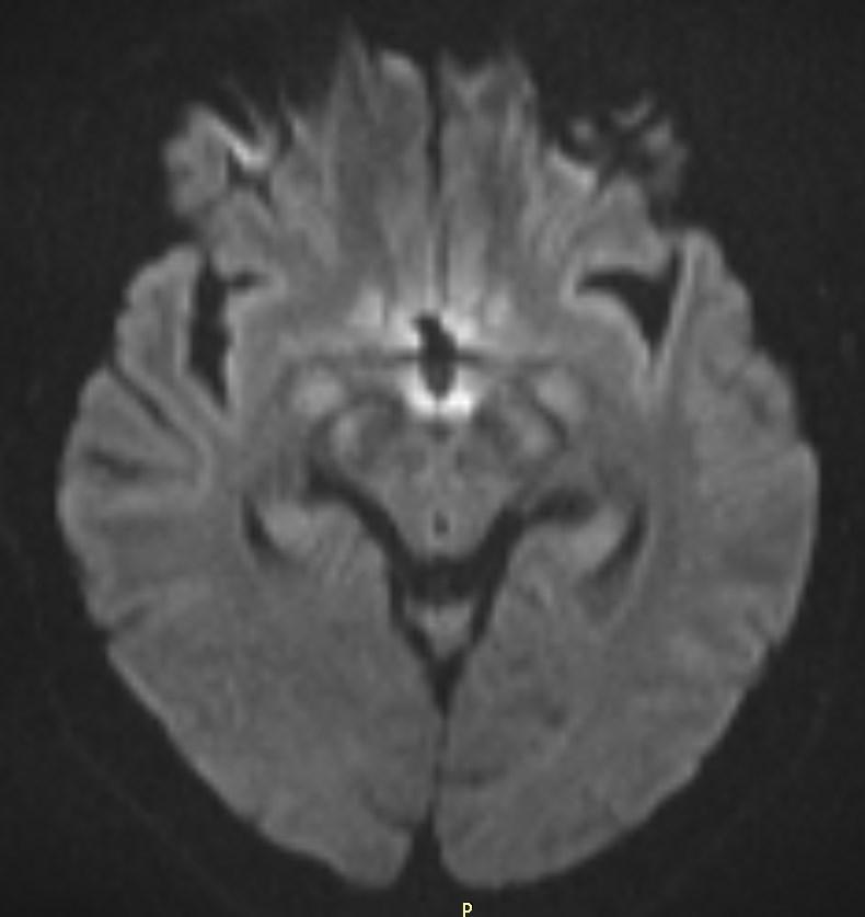
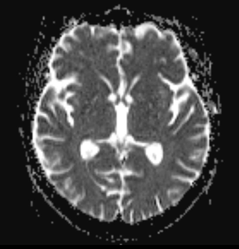
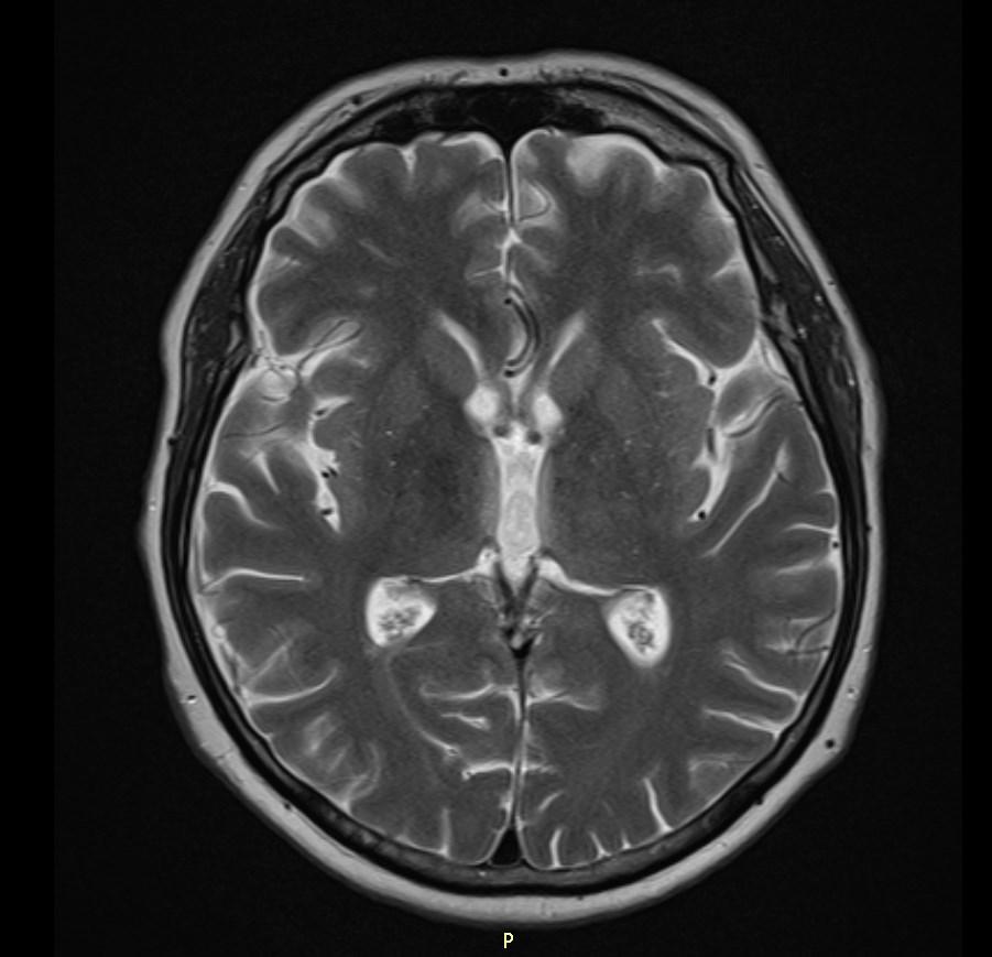
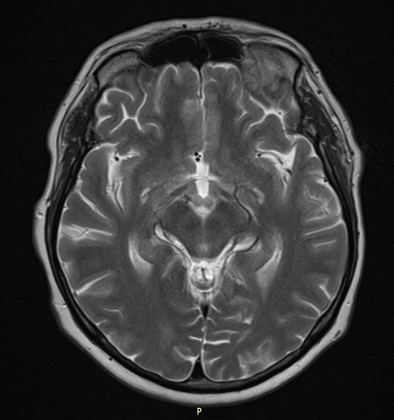
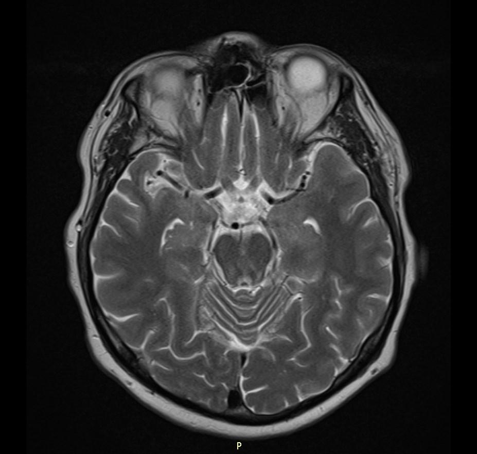
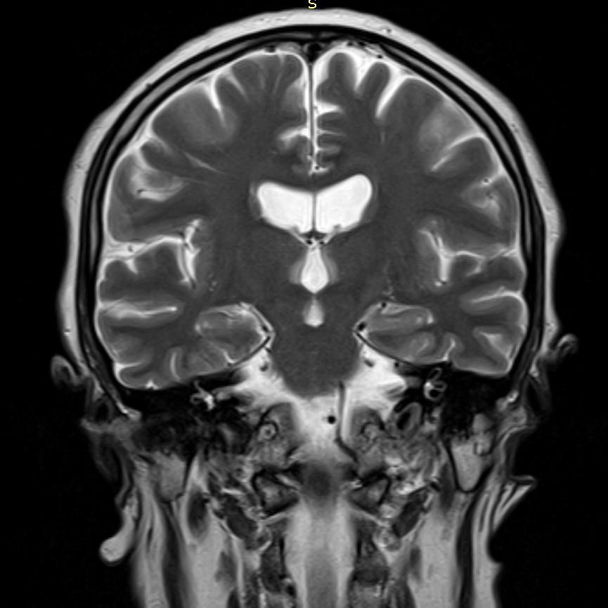
MRI images revealed hyperintensities in the periaqueductal region and the medial thalami.
Wernicke encephalopathy is due to thiamine (vitamin B1) deficiency, and is typically seen in alcoholics.
Clinical presentation
Wernicke encephalopathy was originally described as characterized by the triad of:
Acute confusion
Ataxia
Ophthalmoplegia
Wernicke encephalopathy can evolve into the chronic form of thiamine deficiency known as Korsakoff psychosis, characterised by:
Memory loss (global amnesia)
Confabulation
The two terms are often concatenated to form Wernicke-Korsakoff syndrome.
Etiology
Thiamine deficiency results from malnutrition or malabsorption:
Alcohol abuse
Starvation/fasting
Prolonged total parental nutrition without supplementation
Post bariatric surgery
Hyperemesis gravidum
Gastrointestinal malignancy
Chronic dialysis
MRI
T2/FLAIR: symmetrically increased signal intensity in the mamillary bodies, dorsomedial thalami, tectal plate, periaqueductal area, and around the third ventricle.
References:
1. Zuccoli G, Pipitone N. Neuroimaging findings in acute Wernicke’s encephalopathy: review of the literature. AJR Am J Roentgenol. 2009;192 (2): 501-8.
2. Degnan AJ, Levy LM. Neuroimaging of rapidly progressive dementias, part 2: prion, inflammatory, neoplastic, and other etiologies. AJNR Am J Neuroradiol. 2014;35 (3): 424-31.
3. Thomson AD, Marshall EJ. The natural history and pathophysiology of Wernicke’s Encephalopathy and Korsakoff’s Psychosis. Alcohol Alcohol. 2006;41 (2): 151-8.