*69-year-old male with haematuria and left flank pain.
What is the most likely diagnosis?
Answer
Answer: Transitional cell carcinoma (TCC) of the ureter
Case Discussion:
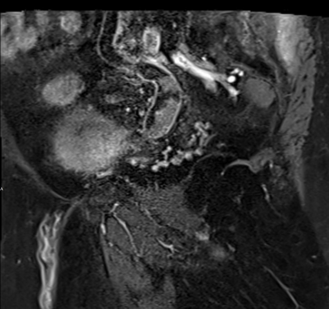
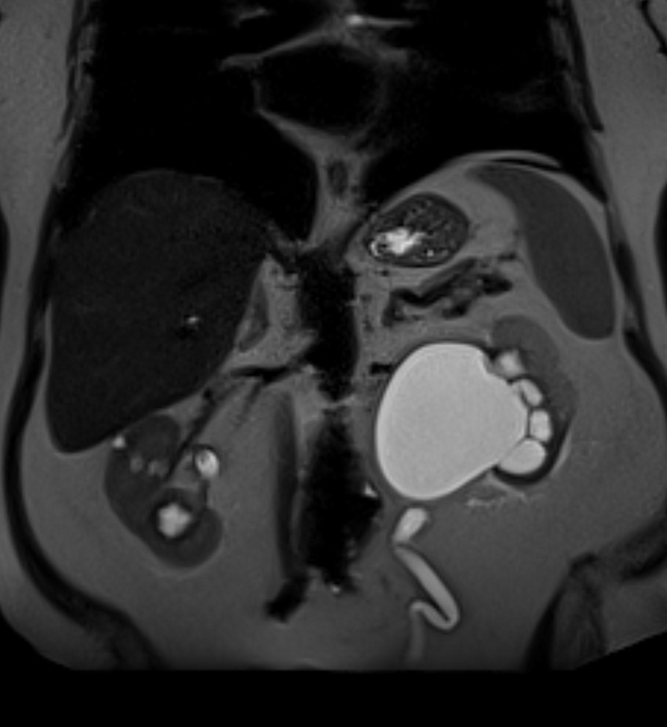
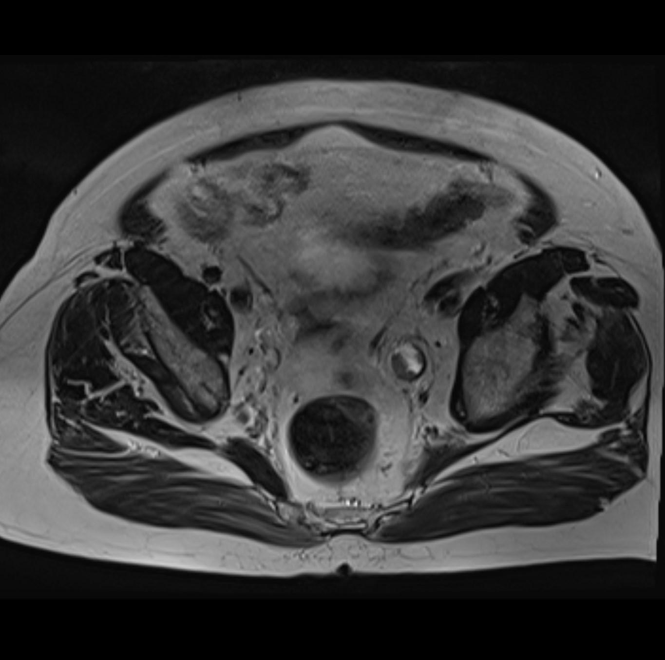
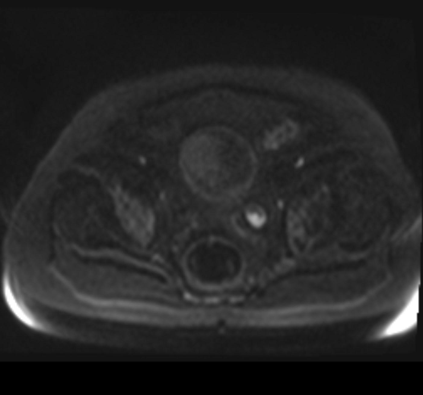
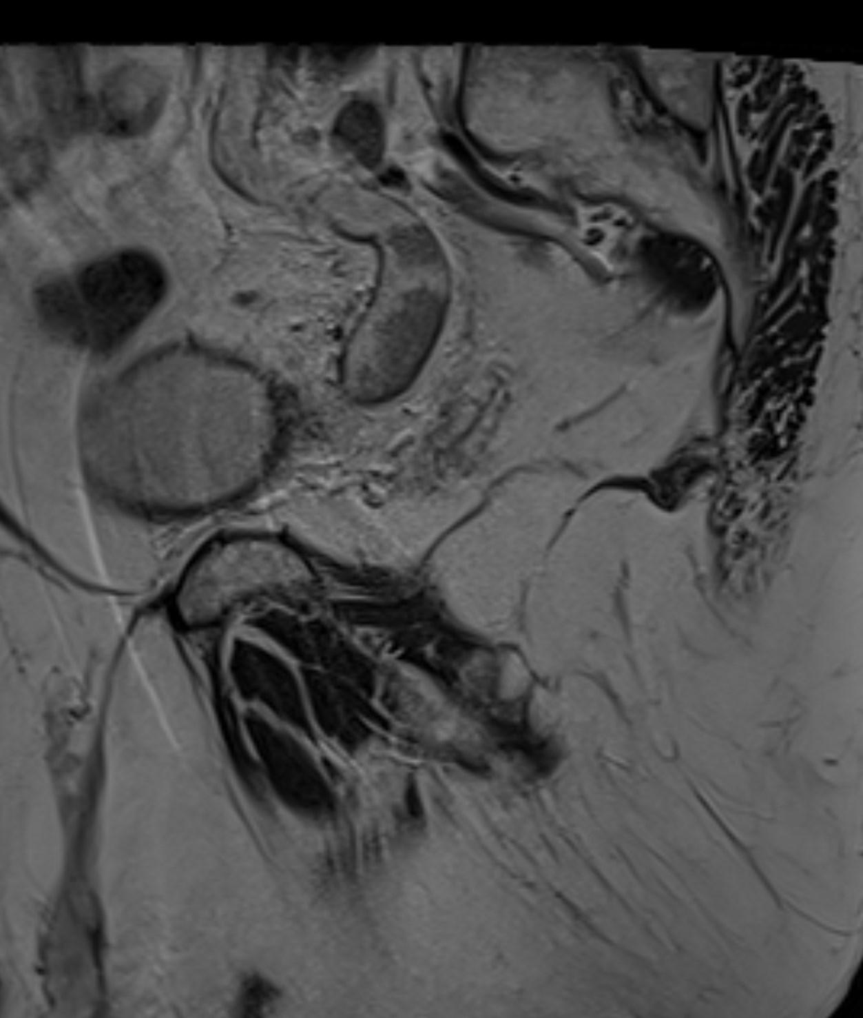
MR images revealed the left dilated ureter and intraluminal polypoidal masses at its distal segment. Associated moderate left hydronephrosis is noted.
TCC of the ureter are rare compared to similar tumors elsewhere along the urinary tract. It accounts for only 1% of all upper urinary tract malignancies.
Location; proximal third: 3%, mid third: 24%, distal third: 73%
References:
1. Leder RA, Dunnick NR. Transitional cell carcinoma of the pelvicalices and ureter. AJR Am J Roentgenol. 1990;155 (4): 713-22.
2. Vikram R, Sandler CM, Ng CS. Imaging and staging of transitional cell carcinoma: part 2, upper urinary tract. AJR Am J Roentgenol. 2009;192 (6): 1488-93.
3. Browne RF, Meehan CP, Colville J et-al. Transitional cell carcinoma of the upper urinary tract: spectrum of imaging findings. Radiographics. 25 (6): 1609-27.