*30-year-old male present with chronic diarrhoea and recurrent abdominal pain.
What is the most likely diagnosis?
Answer
Answer:
Crohn disease
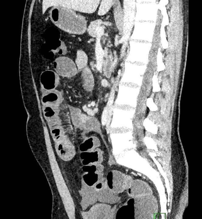
Case Discussion:
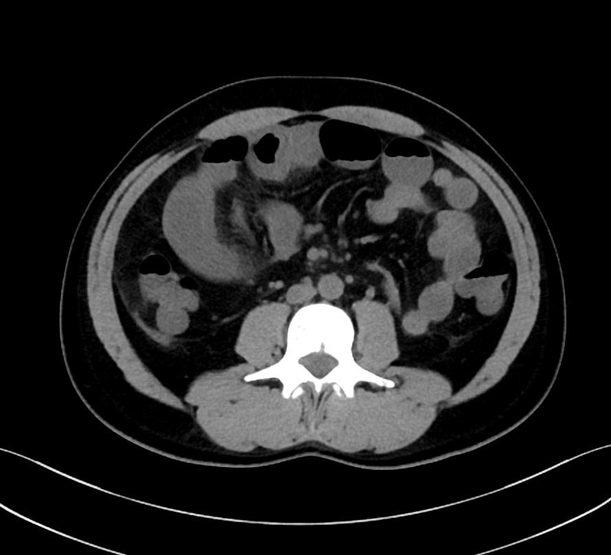
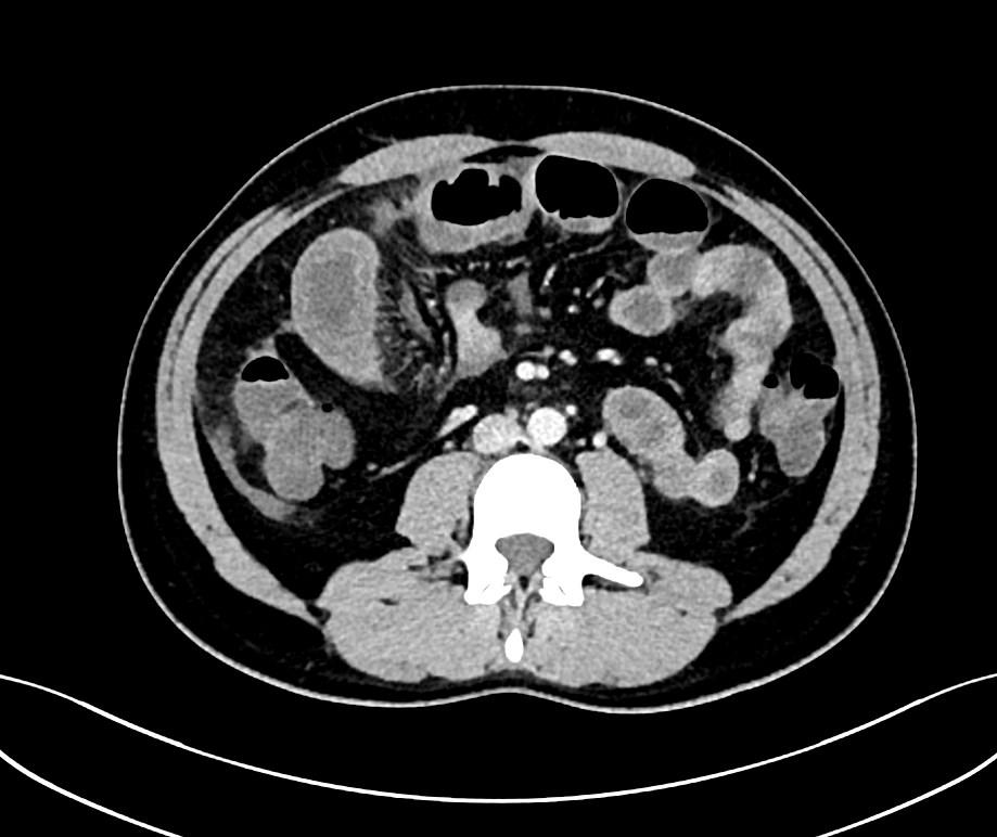
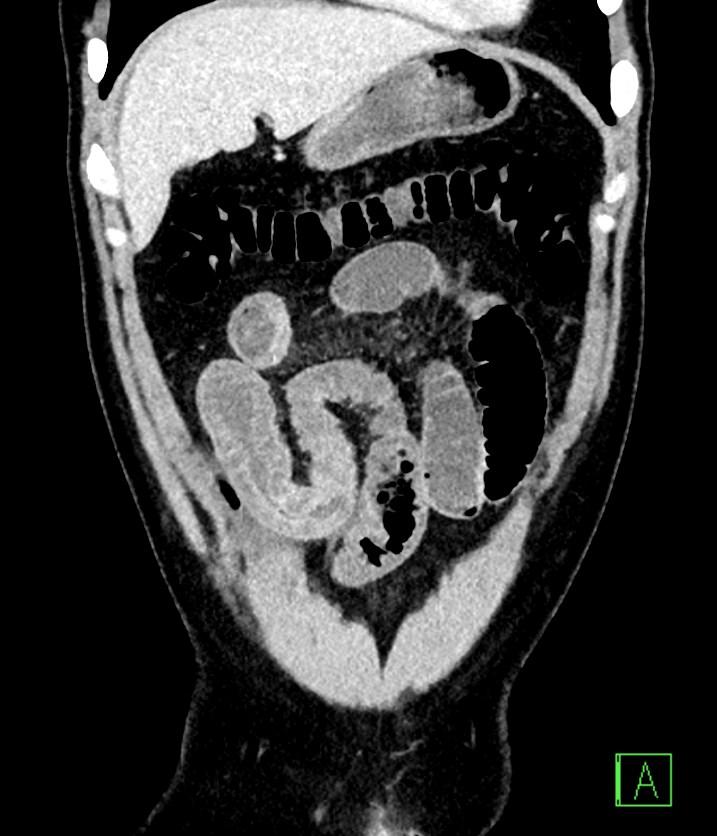
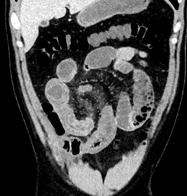
CT images revealed a long segment ileal loop thickening, with prominence of the mesenteric vasculature – comb sign.
Crohn disease is an idiopathic inflammatory bowel disease characterized by widespread discontinuous gastrointestinal tract inflammation. The terminal ileum and proximal colon are most often affected. Extra-intestinal disease is common.
The frequency with which various parts of the gastrointestinal tract are affected varies widely:
small bowel: 70-80%
small and large bowel: 50%
large bowel only: 15-20%
CT and MR enteroclysis are similar in sensitivity for active inflammation. Ultrasound is also an option for diagnosing active disease, follow-up and assessing complications.
CT findings:
fat halo sign
comb sign
bowel wall enhancement
bowel wall thickening
strictures and fistulae
mesenteric/intra-abdominal abscess or phlegmon formation
References:
1. Furukawa A, Saotome T, Yamasaki M et-al. Cross-sectional imaging in Crohn disease. Radiographics. 24 (3): 689-702.
2. Lee SS, Kim AY, Yang SK et-al. Crohn disease of the small bowel: comparison of CT enterography, MR enterography, and small-bowel follow-through as diagnostic techniques. Radiology. 2009;251 (3): 751-61.
3. Gore RM, Balthazar EJ, Ghahremani GG et-al. CT features of ulcerative colitis and Crohn’s disease. AJR Am J Roentgenol. 1996;167 (1): 3-15.
4. Muradali, D. Goldberg, D. US of gastrointestinal tract disease. Radiographics. 2015: 35; 50-70.