*5-year-old girl with hearing loss.


What is the most likely diagnosis?
Answer
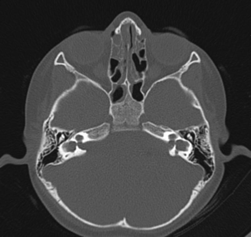
Answer: Bilateral incomplete partition Type I (cystic cochleovestibular anomaly)
Case Discussion:
Axial MR and CT images show the cystic bilateral cochlea (red arrow) and the dilated cystic bilateral vestibule (yellow arrow). The vestibular aqueduct is normal in size.

Cochlear anomalies can be divided as follows:
• complete labyrinthine aplasia (Michel deformity), 3rd week
• cochlear aplasia, 4th week
• common cavity to the cochlea and vestibule, early 5th week
• incomplete partition type I (cystic cochleovestibular anomaly), late 5th week
• cochlear hypoplasia
• incomplete partition type II including Mondini dysplasia ,7th week
References:
1. Head and neck imaging. Ed. by Peter M. Som, Hugh D. Curtin. St Louis (Mo.) : Mosby-Year Book, 2003.
2. Jackler RK, Luxford WM, House WF. Congenital malformations of the inner ear: a classification based on embryogenesis. Laryngoscope. 1987;97 (3 Pt 2 Suppl 40): 2-14.
3. Sennaroglu L, Saatci I. A new classification for cochleovestibular malformations. Laryngoscope. 2002;112 (12): 2230-41.