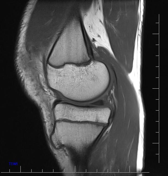
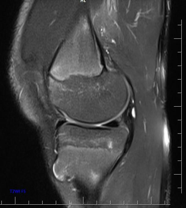
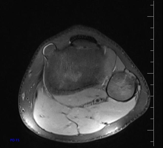
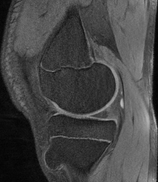
*13-year-old boy with mass on the proximal tibia.
What is the most likely diagnosis?
Answer
Answer: Osteochondroma
Case Discussion:
MR images show osteochondroma on the proximal tibia.

Osteochondromas (also known as osteocartilaginous exostoses) are the most common benign tumors of the bones, accounting for ~35% of all benign tumors of the bones. Osteochondromas are commonly asymptomatic and have a low rate of malignancy if sporadic and solitary. They most commonly develop during the period of rapid skeletal growth. They most commonly arise from appendicular skeleton, especially around the knee.
Types
• Solitary Osetocartilaginous Exostosis (most common)
• Hereditary Multiple Exostoses (also known as diaphyseal aclasis)
Radiographic features
They can be either pedunculated or sessile, and are seen in the metaphyseal region typically projecting away from the epiphysis on plain film.
CT demonstrates cortical, medullary continuity and the cartilage cap.
MRI is the best imaging procedure to evaluate cartilage thickness, presence of edema in bone or adjacent soft tissues, and visualizing neurovascular structures in the vicinity.
The cartilage cap appears the same as cartilage elsewhere, with intermediate to low signal on T1WI and high signal on T2WI. The cartilage cap of over 1.5 cm in thickness is suspicious for malignant degeneration. The cartilaginous cap itself should not enhance.
References:
1. Dähnert W. Radiology Review Manual. Lippincott Williams & Wilkins. 2007
2. Eisenberg RL, Johnson NM. Comprehensive radiographic pathology. Mosby Inc. 2007
3. Murphey MD, Choi JJ, Kransdorf MJ et-al. Imaging of osteochondroma: variants and complications with radiologic-pathologic correlation. Radiographics. 20 (5): 1407-34.