*23-year-old female with headache.
What is the most likely diagnosis?
Answer
Answer: Cavum velum interpositum
Case Discussion:
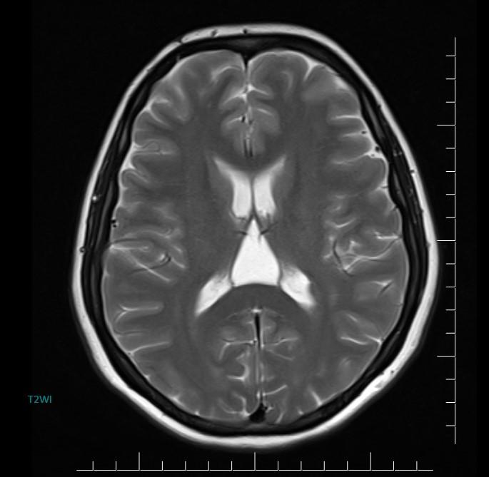
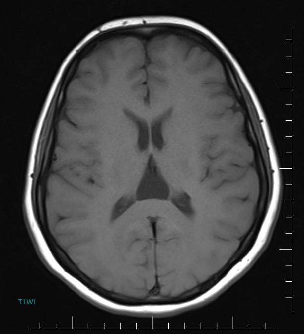
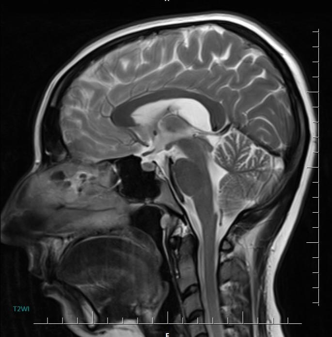
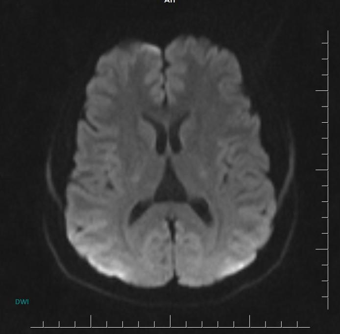
MR images incidentally revealed a triangular-shaped CSF space between the lateral ventricles (arrows).

In the brain, the cavum velum interpositum is a condition where there is a dilated cerebrospinal fluid (CSF) space involving the cistern of the velum interpositum. It usually occurs in newborns.
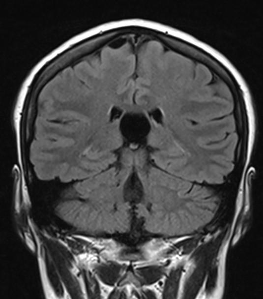
Axial MR/CT image shows a triangular-shaped CSF space situated behind the foramen of Monro, between the lateral ventricles. Coronal image demonstrates the elevated and splayed fornices.
There are usually no associated anomalies, though larger lesions can cause an obstructive hydrocephalus. Treatment is usually unnecessary.
References:
1 .Epelman M, Daneman A, Blaser SI et-al. Differential diagnosis of intracranial cystic lesions at head US: correlation with CT and MR imaging. Radiographics. 26 (1): 173-96.
2. L. M. Ketonen. [et al.]. Pediatric Brain and Spine. Springer. 2005