*28 day-old female newborn


What is the most likely diagnosis?
Answer
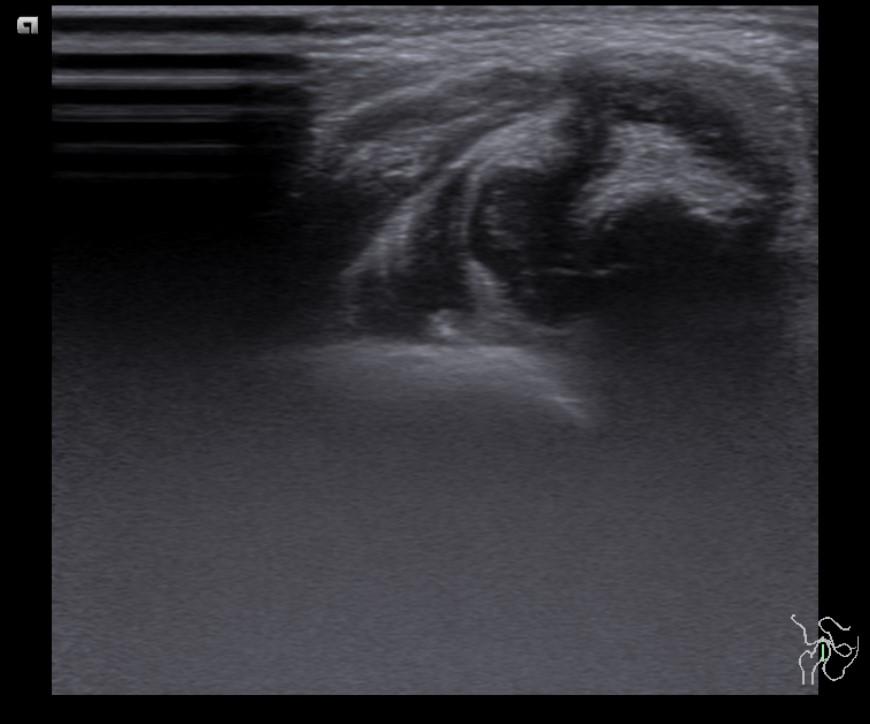
Answer: Developmental dysplasia of the hip
Case Discussion:
Coronal ultrasound images demonstrated elevated labrum, dislocation of femoral head, and hypoplastic acetabular fossa (type IV according to Graf).
Developmental dysplasia of the hip is a spectrum of disorders that leads to hip subluxation and dislocation. Ultrasound is the modality of choice in the infant prior to ossification of the proximal femoral epiphysis (<6 months). The reported incidence of DDH in infants varies between 1.5 and 20 per 1000 births. Risk factors include: female gender (M:F ratio ~1:8), family history, breech presentation, oligohydramnios (1, 2).
References:
1. US Preventive Services Task Force. “Screening for Developmental Dysplasia of the Hip: Recommendation Statement.” Pediatrics 117, no. 3 (3, 2006): 898-902.
2. Taeusch H. William, Roberta A. Ballard, Christine A. Gleason and Mary Ellen Avery. Avery’s diseases of the newborn. Elsevier Health Sciences, 2005