**A 61-year-old woman presented with headache, confusion and neurologic deficits


What is the most likely diagnosis?
1. Developmental venous anomaly
2. Subdural hematoma
3. Diffuse axonal injury
4. Cerebral Vein and Sinus Trombosis
Answer
Case Discussion:
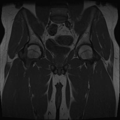
MRI and MR venography showed subacute thrombosis of all dural sinuses and some vein. MRI demonstrated right frontal, parietal, and left parietal focal subcortical parenchymal haemorrhages. CT demonstrated empty delta sign.
Cerebral vein and sinus thrombosis (CVT) is a disorder that results wide variety of clinical manifestations. It is a rare condition compared with arterial stroke and generally occurs in young females (1). Oral contraceptive pills, pregnancy, steroids, prothrombotic conditions, trauma, dehydration and sepsis are well known risk factors (1). CT venogram shows ‘empty delta sign’ in the sagittal sinus as filling defect (2). 50% of cases CVT progresses to venous infarction (2). In the acute stage of thrombus development (0–5 days), the signal is mostly isointense on T1WI and hypointense on T2WI due to deoxyhemoglobin in red blood cells in the thrombus (4). In the subacute stage of thrombus formation (6–15 days), the signal is primarily hyperintense on both T1WI and T2WI due to methemoglobin within the thrombus (5). In the chronic stage of thrombus formation (>15 day), characteristically isointense on T1WI and isointense/hyperintense on T2WI, compared with the MR signal in normal brain parenchyma (6,7). Treatment is heparin and in selected cases local endovascular thrombolysis may help (1).
References:
1.Ferro JM, Canhão P, Stam J et-al. Prognosis of cerebral vein and dural sinus thrombosis: results of the International Study on Cerebral Vein and Dural Sinus Thrombosis (ISCVT). Stroke. 2004;35 (3): 664-70.
2. Rodallec MH, Krainik A, Feydy A et-al. Cerebral venous thrombosis and multidetector CT angiography: tips and tricks. Radiographics. 2006;26 Suppl 1 : S5-18.
4. Hinman JM, Provenzale JM. Hypointense thrombus on T2-weighted MR imaging: a potential pitfall in the diagnosis of dural sinus thrombosis. Eur J Radiol 2002; 41:147–152.
5. Dormont D, Anxionnat R, Evrard S, Louaille C, Chiras J, Marsault C. MRI in cerebral venous thrombosis. J Neuroradiol 1994; 21(2):81–99.
6. Selim M, Fink J, Linfante I, Kumar S, Schlaug G, Caplan LR. Diagnosis of cerebral venous thrombosis with echo-planar T2*-weighted magnetic resonance imaging. Arch Neurol 2002; 59:1021–1026.
7. Isensee C, Reul J, Thron D. Magnetic resonance imaging of thrombosed dural sinuses. Stroke 1994; 25:29–34.