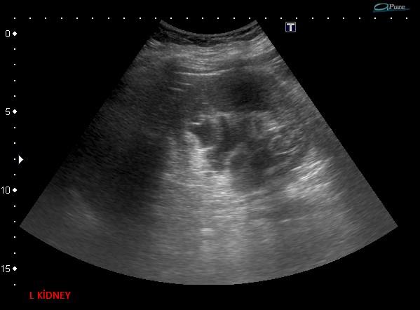
*66 year old female with left flank pain.


What is the most likely diagnosis?
1. Hereditary polycystic kidney disease
2. Familial renal lymphangiomatosis
3. Cystic renal dysplasia
4. Renal lymphangiectasia
Answer
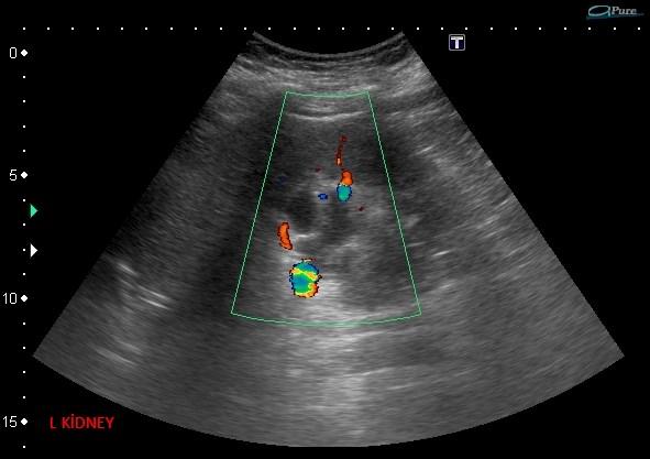
Answer: Renal lymphangiectasia
Case Discussion:
Abdominal US images and axial CT sections demonstrating bilateral renal lymphangiectasia. Axial CT section in nephrographic phase and axial and coronal sections during the excretory phase showing bilateral cystic lesions of the renal sinus as well as the stone in the left distal ureter (arrow). This causes extrinsic compression and displacement of the adjacent collecting system.
Renal lymphangiectasia (also known as a renal lymphangioma) is an uncommon condition characterized by different degrees of dilatation of the lymphatic ducts. It is caused by an abnormal development of the lymphatic ducts, with single or multilocular fluid-filled cavities. It is thought to be because of an abnormal communication between the renal lymphatics and the wider retroperitoneal lymphatics.
References:
1. Ashraf K, Raza SS, Ashraf O, et al. Renal lymphangiectasia. Br J Radiol. 2007;80:117- 8.